Introduction:
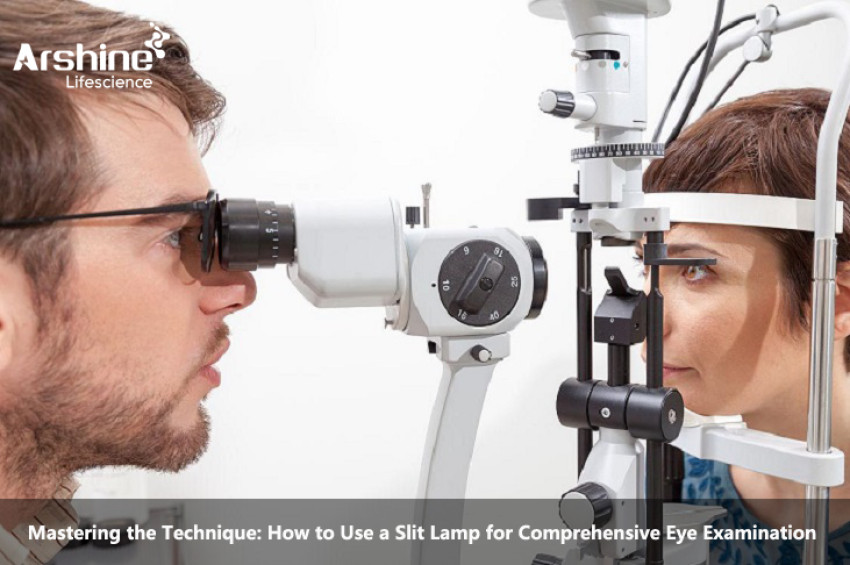
A slit lamp is a powerful diagnostic instrument used by ophthalmologists and optometrists to perform detailed examinations of the eye's anterior segment. This sophisticated device provides a magnified and illuminated view of the cornea, iris, lens, and other anterior eye structures. Mastering the technique of using a slit lamp is essential for accurate diagnosis and effective management of various eye conditions. In this comprehensive guide, we will explore step-by-step instructions on how to use a slit lamp for a comprehensive eye examination.
A slit lamp combines a high-intensity light source with a binocular microscope and a variable-width slit beam. The light source, typically a halogen or LED bulb, emits a narrow beam of light, which can be adjusted in width and direction. The microscope provides a magnified view of the anterior segment, allowing detailed examination of the eye's structures.
The slit lamp also includes filters, such as a cobalt blue filter and a yellow filter, which aid in assessing different eye conditions, including corneal abrasions, cataracts, and dry eye syndrome.
1.Patient Preparation:
Before using the slit lamp, ensure the patient is comfortably seated and properly positioned. Adjust the height and angle of the slit lamp to match the patient's eye level. Dim the room lights to improve visualization and minimize glare during the examination.
Explain the procedure to the patient, addressing any concerns they may have. Reassure them that the examination is painless and non-invasive.
2.Slit Beam Alignment:
Start by aligning the slit beam in the vertical position. Adjust the width of the slit beam to about 3-5mm to cover the entire cornea and iris. Focus the microscope using the ocular focusing wheel until the patient's eye comes into sharp view.
Position the patient's forehead against the headrest and ask them to rest their chin on the chin rest. Align the patient's eye with the microscope's eyepiece to ensure a clear view of the eye's structures.
3.External Eye Examination:
Begin the examination by assessing the external eye structures. Move the slit beam horizontally across the eye's surface while observing the eyelids, eyelashes, conjunctiva, and sclera. Look for signs of inflammation, swelling, redness, or any abnormalities.
4.Corneal Examination:
Next, focus the slit beam on the cornea, the clear front surface of the eye. Examine the cornea for any signs of abrasions, ulcers, infections, or irregularities in its shape. The cobalt blue filter can be used to detect corneal staining or the presence of foreign bodies.
Adjust the slit beam to an oblique angle to visualize the corneal layers and assess for signs of edema or dystrophies.
5.Anterior Chamber Examination:
Move the slit beam slightly downward to examine the anterior chamber, the fluid-filled space between the cornea and the iris. Observe the depth and clarity of the anterior chamber, looking for signs of inflammation, cells, flare, or abnormal fluid accumulation.
6.Iris Examination:
Continue moving the slit beam downward to focus on the iris, the colored part of the eye. Examine the iris for any abnormalities, such as irregularities in its structure, pigment dispersion, or signs of inflammation.
7.Lens Examination:
Finally, adjust the slit beam to the horizontal position and move it further downward to visualize the lens, located behind the iris. Examine the lens for signs of cataracts, opacities, or any structural changes that could affect vision.
8.Pupil Examination:
During the examination, assess the size and reactivity of the patient's pupils using the slit lamp. Observe the pupils' response to light and accommodation, looking for any signs of abnormal pupil reactions.
9.Filter Application:
As necessary, apply the appropriate filters, such as the cobalt blue filter or the yellow filter, to enhance the examination of specific eye conditions. For example, the cobalt blue filter can be used to detect corneal abrasions or ulcers, while the yellow filter helps assess cataracts or macular changes.
10.Documentation and Communication:
Throughout the examination, record any significant findings in the patient's medical record. If any abnormalities or conditions are detected, communicate the findings with the patient and discuss the appropriate management plan.
Conclusion:
The slit lamp is an indispensable tool in comprehensive eye examination, providing a magnified and illuminated view of the eye's anterior segment. By mastering the technique of using a slit lamp, ophthalmologists and optometrists can accurately diagnose and manage various eye conditions, ranging from corneal abrasions and cataracts to iritis and anterior chamber abnormalities.
A systematic approach to the examination, combined with proper patient positioning and communication, ensures a thorough and comfortable experience for the patient while yielding valuable diagnostic information for the eye care provider.
Regular use of the slit lamp in clinical practice enhances the precision of eye examinations, leading to better patient outcomes and improved management of eye conditions. As technology continues to advance, the slit lamp's capabilities may expand further, contributing to the ongoing advancements in eye care and vision health.
https://www.arshinemedical.com/Industry-information/mastering-the-technique-how-to-use-a-slit-lamp-for-comprehensive-eye-examination